

![]()
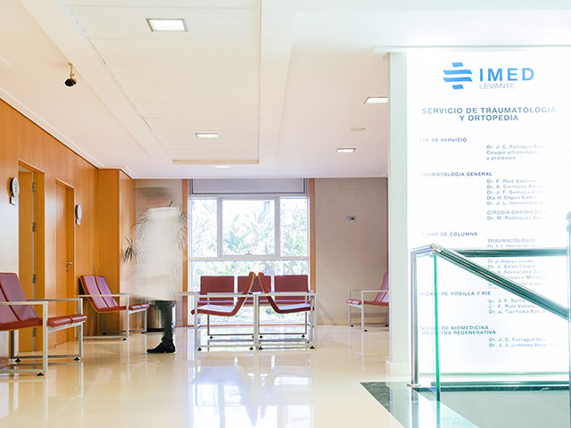
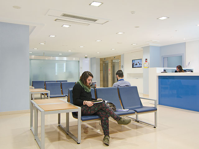
With the confidence and security of a hospital environment
Fertility Clinics and Reproductive Medicine
At VITA, the first diagnostic consultation is completely free of charge and without obligation. In this first visit we will evaluate all aspects related to your fertility and that of your partner, studying the most appropriate treatment for your case.
In vitro fertilization (IVF) is the most widely used assisted reproduction technique, achieving pregnancy rates much higher than other techniques such as artificial insemination.
Anonymous egg donation is the most effective assisted reproduction technique, achieving the best pregnancy rates.
One of the simplest techniques
Shared maternity technique for female couples.
It successfully solves most male infertility problems.
Synchronises ovulation with sexual intercourse.
Egg freezing to preserve fertility.
Reception of previously vitrified donated embryos.
Your first free medical appointment
Request a free appointment with a specialist in reproductive medicine so that you can solve your doubts.
VITA Reproductive Medicine centers are specialized in fertility and assisted reproduction treatments. They have a multidisciplinary team made up of prestigious professionals from gynecologists, embryologists, geneticists, urologists, nurses, anesthesiologists, psychologists to patient assistants who will accompany you on the road to motherhood.
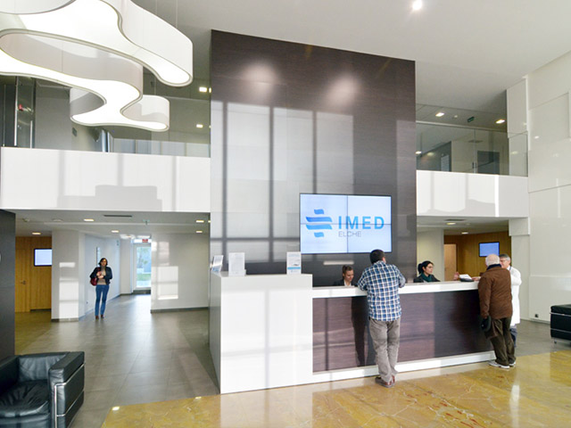
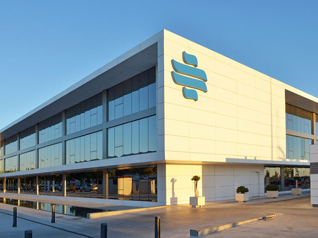
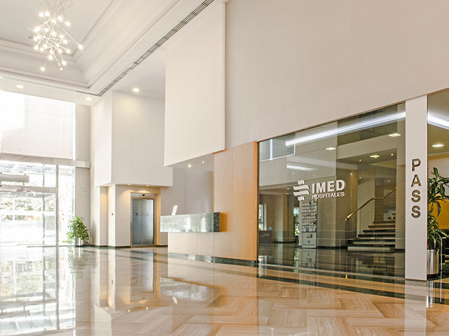
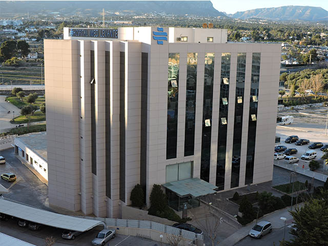
VITA centers, located in Elche, Benidorm, Gandia and Alcoy, are integrated within the hospital setting of IMED Hospitales, with all the advantages that this implies for your health and safety during treatments.
In VITA we take the greatest care over all the details and we treat all our patients in a totally personalized way, with the warmth and naturalness they deserve.
A complete fertility study that will enable you to know the possibilities you have to get pregnant, now or in the future.
VITA, centres specialising in fertility, assisted reproduction and reproductive medicine, within the IMED Hospitales facilities.
Age plays a key role in getting pregnant. Check the chances of pregnancy according to your age.
Meet the VITA team, made up of a multidisciplinary team of recognised prestige.
Experiencias de madres que consiguieron su sueño de ser madres gracias los tratamientos de fertilidad y reproducción asistida de VITA.
Many women are unable to get pregnant with their own eggs and need the help of an egg donor to fulfil their dream.
WHY CHOOSE VITA REPRODUCTIVE MEDICINE?
Discover why VITA is a reference in fertility treatments in Alicante

- Hospital Environment VITA clinics are located within the IMED Elche and Levante Hospitals in Benidorm, reference hospitals in Alicante..
- We insure your pregnancy At VITA we offer the possibility of contracting our “BONOFIV”. We will refund the cost paid if you do not get pregnant after 4 cycles of egg donation.
- Agreements with most insurance companies VITA has agreements with the majority of insurance companies and other collectives that allow you to access special conditions.
- Personalised attention throughout the treatment Throughout the treatment you will be accompanied by a VITA specialist who will advise you and answer all your questions.
- Excellent success rates Treatments offered by VITA has above average success rates, with excellent pregnancy results.
- No waiting lists and flexible schedules Start your treatment whenever you want, with flexible timetables 24 hours a day when you start the process. At VITA we adapt to your needs.
- Tailored financing In addition to reasonable prices, VITA offers you the possibility of financing your treatment in easy instalments, with an immediate study and processing and without changing banks.
- Access to online medical records VITA patients can access their medical records online, and have the possibility to make medical consultations via Skype..


ASSISTED REPRODUCTION AND REPRODUCTIVE MEDICINE
News and novelties on assisted reproduction treatment, techniques and tests.
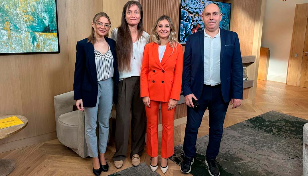
VITA brings its fertility treatments to british patients
The VITA team recently visited London with a clear objective: to bring our fertility treatments to british women who want to become mothers and raise awareness of them. Through informative meetings and personalised consultations, attendees [...]
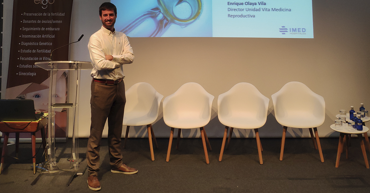
Dr. Enrique Olaya, VITA Reproductive Medicine Manager, speaker on the I Fertility and Assisted Reproduction Workshop.
The first edition of the Fertility and Assisted Reproduction Workshop was hold in Gijon the 18th and 19th of October. This event that took place in Laboral Ciudad de la Cultura in Gijon, was directed [...]
Vita, the only center with his own laboratory in the Marina Baixa
One of the main reasons why many of our patients choose Vita and why we are the reference center in fertility treatments in the province of Alicante is because we have our own laboratory in [...]